
Figure 5.
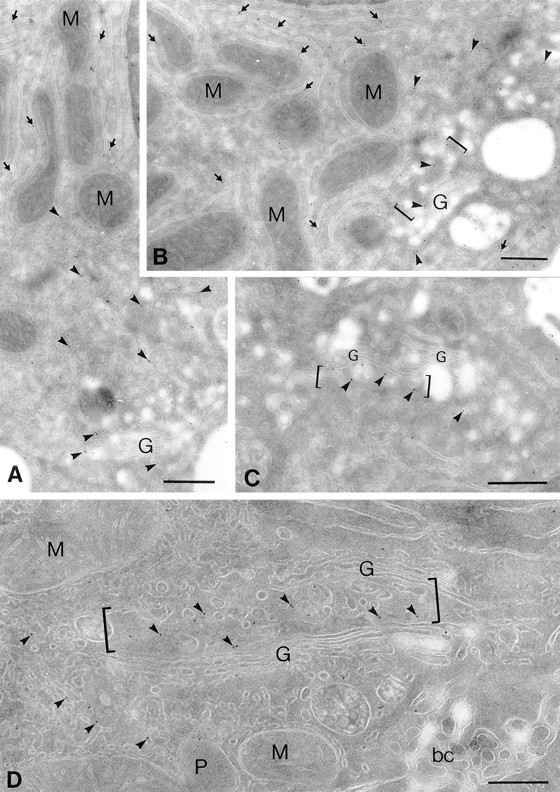
In situ distribution of the α2 protein in rat liver hepatocyte ultrathin cryosections. Cryosections of rat liver hepatocytes were immunolabeled with the rabbit anti-C–terminus antibody against the α2 sequence followed by goat anti– rabbit IgG 10-nm-gold conjugates. Representative profiles of labeling within secretory compartments in liver hepatocytes are shown. Profiles of flattened cisternae of the rough ER are labeled along their length (A and B, arrows); note the gold particles lining mostly the cytosolic surface of the rough ER cisternae where the COOH-terminal domain of the α2 sequence is expected. α2 labeling is distributed throughout tubular smooth membranous profiles (arrowheads) in the Golgi/bile canalicular region of hepatocytes; prominent gold particle labeling can be seen in tubulovesicular membranous profiles closely approaching one side of a given Golgi apparatus (B–D, brackets). Labeling within the Golgi apparatus for the α2 protein is similarly restricted to one of the saccules on one side of a Golgi stack, whereas most other saccules reveal negligible labeling. Mitochondria (M), peroxisomes (P), and bile canaliculus (bc) were largely devoid of labeling. Bars, 400 nm.