
Figure 9.
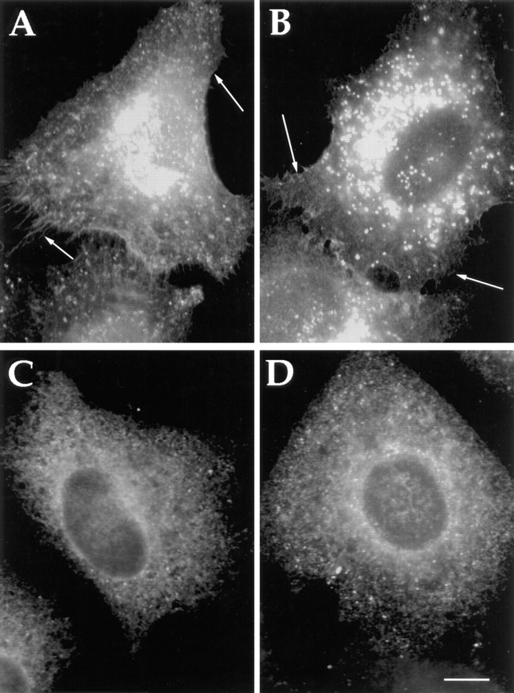
Altered distribution of family members after mutation of K(X)KK(X) on FF motifs of the cytosolic domains of α2 and β1 proteins. Cotransfection of all five p24s in where either the KK (A and B) or the FF (C and D) motif has been altered to SS or AA, respectively, in both α2 and δ1. Altered redistribution and apparent cell surface staining can be seen with the KK/SS mutants (shown is α2)(A). Remarkably, the unmutated members (shown is γ3/4) (B) also reveals redistribution to the cell surface. Similarly, an apparent ER staining can be seen with the FF/AA mutants (shown is α2)(C). This also leads to the apparent redistribution of the unmutated members to the ER (shown is γ3/4)(D). Bar, 5 μm.