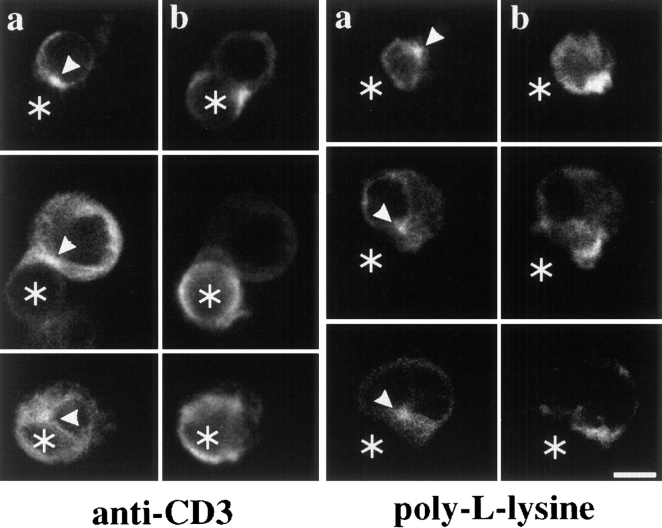
Figure 1.
Anti–TCR-coated latex beads induce MTOC reorientation and polarized actin polymerization in Jurkat T cells. Jurkat T cells were mixed at a 1:2 ratio with anti–CD3ε-coated latex beads (left) or poly-l-lysine-coated latex beads (right). After 30 min at 37°C, conjugates were stained with the antitubulin antibody YOL1/34 (a) and rhodamine-phalloidin to visualize F-actin (b). The position of cell-bound latex beads is indicated by an asterisk, the position of the MTOC by an arrowhead. Bar, 5 μm.

