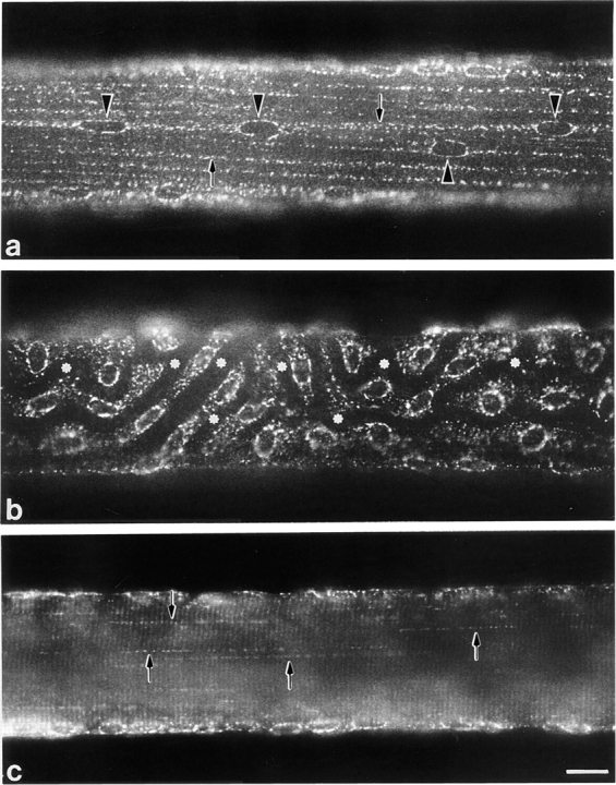
Figure 1.

Immunofluorescence localization of GLUT4 in single fibers from basal soleus muscle. Single fibers were stained with anti-GLUT4 antiserum followed by biotin-conjugated anti-rabbit IgG and FITC-labeled streptavidin. (a) At the surface of the fibers, in some areas, myonuclei (arrowheads) are aligned with the axis of the fiber and joined by regular rows of GLUT4 aggregates (arrows). (b) In other areas, nuclei are not aligned with the axis of the fiber and the staining pattern appears less orderly. Dark channels (asterisks) correspond to capillaries. (c) In the core of the fibers, the staining consists of dotted lines (arrows) and weak cross-striations. Bar, 10 μm.
