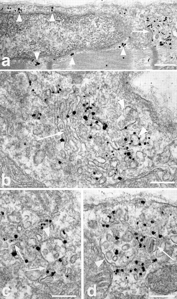
Figure 12.

Immunogold EM localization of the TfR in basal fibers. Single fibers were stained for TfR and embedded in epoxy resin as described in Materials and Methods. (a) Low magnification overview showing an area around a nucleus near the plasma membrane. TfR is present in large aggregates in the area of the Golgi complex (arrow) and in smaller aggregates all around the nucleus (arrowheads) in a pattern that resembles that seen for GLUT4 in Fig. 4. (b) A stack of Golgi cisternae (arrow) showing TfR labeling, both in the cisternae and in vesicles of different sizes around the cisternae (arrowheads). (c and d) Labeling is often seen associated with multivesicular bodies (arrowheads) of different sizes. In c, the labeling seems excluded from the internal structures whereas in d they are labeled. Note also a network of labeled, tubulovesicular structures (arrows) around the multivesicular bodies. Bars: (a) 0.5 μm; (b–d) 0.2 μm.
