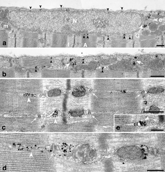
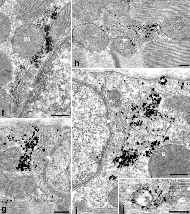
Figure 4.


Localization of GLUT4 in basal fibers observed by preembedding immunogold electron microscopy. Single fibers were stained and embedded in epoxy resin as described in Materials and Methods. (a and b) Low magnification views of two areas close to the plasma membrane, one with a nucleus (a), the other with myofibrils extending very close to the plasma membrane (b). Both show small (arrowheads) and large (arrow) clusters of GLUT4. In a they surround a nucleus (N). The large clusters are next to a Golgi complex (G) and to multivesicular bodies (e; see enlarged area in i). Other recognizable elements are mitochondria (m) and myofibrils that fill the core of the fiber, with the characteristic A and I band alternation. Note the almost complete absence of single grains on the plasma membrane. (c– e) Distribution of GLUT4 in the core of the fibers. In basal conditions, most GLUT4 staining is in clusters of grains (arrowheads) in the I band, near the A–I band intersection, or more rarely in the A band. T tubules at the triad junctions (small arrows) are unlabeled but there is labeling of terminal cisternae of the SR (d and e) and of what appears to be the fenestrated SR in the A band (arrowheads). (f–j) Details of GLUT4 localization showing association with Golgi complex and endosomes. (f–g) Next to nuclei (N) that are close to the fiber surface, stacks of Golgi cisternae (arrowheads) can be recognized. GLUT4 is concentrated in the cisternae farthest away from the nucleus and in tubulovesicular structures just beyond or lateral to the cisternae. In h, GLUT4 is associated with a Golgi complex which is in the myofibrillar core and is not associated with a nucleus. In i, the gold grains are next to Golgi cisternae and to mutivesicular bodies, presumably endosomes (e), whereas j shows an abundantly labeled endosome in the myofibrillar core. Bars: (a–c) 0.5 μm; (d–j) 0.2 μm.
