Figure 7.


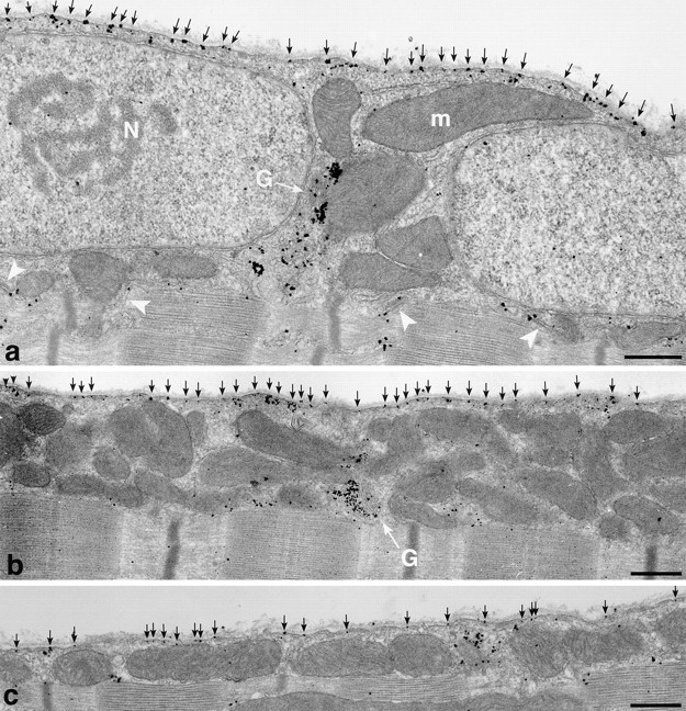
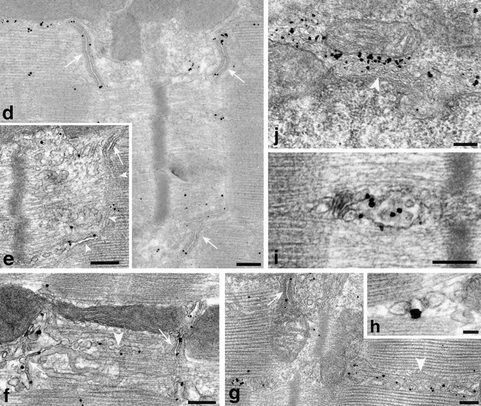
Translocation of GLUT4 in stimulated fibers observed by preembedding immunogold electron microscopy. (a) Overview of a fiber that has been maximally stimulated with both insulin and contractions, showing two nuclei (N) and several mitochondria (m). The plasma membrane contains numerous single grains (black arrows) and the T tubules of several triad junctions also appear labeled (white arrowheads). GLUT4 labeling is also found at the pole of one of the nuclei, in the Golgi complex region (G). (b) Despite the heavy labeling of the plasma membrane (black arrows) in a fiber from contraction-stimulated muscle, the Golgi stack (G) still has fairly dense polarized GLUT4 labeling. (c) Overview of a fiber from an insulin-stimulated muscle showing dense labeling of the plasma membrane (black arrows). (d–h) Labeling of junctional T tubules (white arrows) in fibers from exercise- (d and f) or insulin and exercise-stimulated (e and g) muscle. In e, the nonjunctional part of a T tubule leaving a triad can be seen (white arrowheads) coursing between the myofibrils. The labeling of apparently fenestrated SR (f and g, white arrowheads) is mostly in the form of single grains and not of clusters of grains as seen in nonstimulated fibers. (h) Labeling of the T tubule in a cross-sectioned triad in a fiber from insulin-stimulated muscle. (i) Labeling of an endosome in the myofibrillar core of an insulin-stimulated fiber. (j) The labeling of the TGN area in an insulin and exercise-stimulated fiber appears lighter than in basal fibers. Bars: (a–c) 0.5 μm; (d–g, i, and j) 0.2 μm; (h) 0.05 μm.
