
Figure 6.
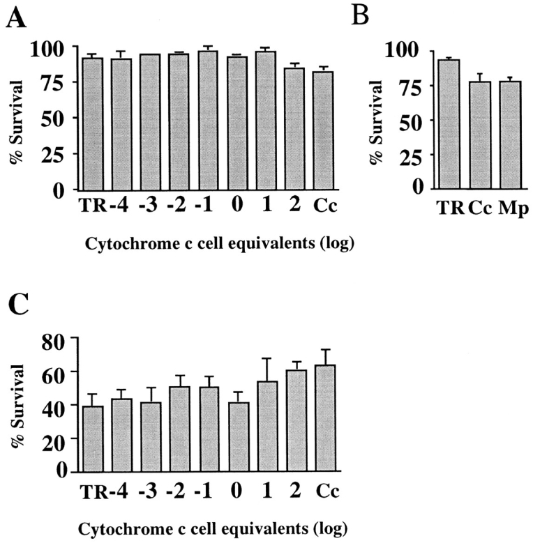
Microinjection of cytochrome c does not induce or accelerate apoptosis in SCG neurons. SCG neurons were microinjected with cytochrome c and counted 2–4 h later. (A) Cells were maintained in NGF for 48 h before counting the surviving cells. The amount of cytochrome c injected is shown as log10 multiples of 1 cell equivalent (70 μg/ml in needle), except for lane TR, which contained no cytochrome c, and lane Cc, in which 17.5 mg/ml of cytochrome c was used. (B) Cells were maintained in NGF for 72 h before counting surviving cells. The microinjection mix in lane TR contained no cytochrome c, 1.45 mM cytochrome c (17.5 mg/ml) in lane Cc, and 1.45 mM microperoxidase in lane Mp. (C) Cells were withdrawn from NGF for 48 h before counting the surviving cells. The amount of cytochrome c injected is shown as log10 multiples of 1 cell equivalent (70 μg/ml in needle), except for lane TR, which contained no cytochrome c, and lane Cc, in which 17.5 mg/ml of cytochrome c was used. The results are expressed as a percentage of the cells initially surviving injection. 150–200 cells were injected per coverslip, and the results shown are the average of three to four experiments. The error bars represent SEM.