
Figure 7.
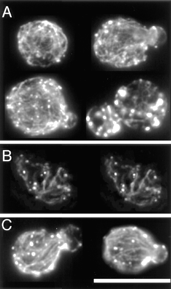
The actin cytoskeleton of the bee1/las17 mutant. (A) Single–focal plane images of four cells at different stages of the cell cycle with a completely random cable meshwork. (B) A stereopair of another such cell. Movie 3 shows rotating images of this cell, with 180 frames at sequential projections of 2° angles. Movies are available at www.cooperlab.wustl.edu. (C) Single– focal plane images of two cells with partial orientation of cables. Cells were stained with rhodamine-phalloidin. Deconvolution was with the EM method. Bar, 10 μm.