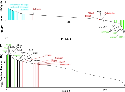
Fig. 5.
ER proteins on LBVs are integral to the membrane but constitute only a small fraction of the total ER. (a) Ranked relative abundances of proteins on LBVs isolated from RAW cells at pH 11.5 versus at pH 7.2. Specific ribosomal proteins (blue), ER markers (red), integral LBV membrane proteins (red), and membrane-associated proteins (green) are indicated. PDIA3, protein disulfide isomerase 3; αSNAP, soluble NSF attachment protein; FcγR, Fcγ receptor; CD-M6PR, cation-dependent mannose 6-phosphate receptor. (b) The amount of each protein found on LBVs at 10 min, expressed as a fraction of the total amount of that protein in the cell normalized for the fraction of cells taking up beads. Specific ER markers (red), endosomal/phagosomal markers (black), and plasma membrane markers (green) are indicated. TLR7, Toll-like receptor 7; Cath, cathepsin; Na/Kβ3, Na+/K+-ATPase β3 subunit. The ratios are cut off at 100-fold up or down because the linearity of the signal in the Orbitrap starts to drop off beyond this.

