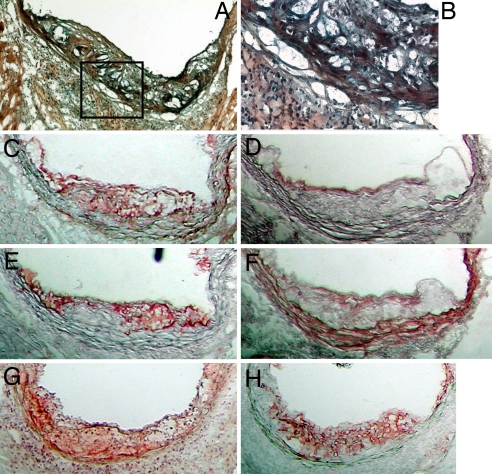
Fig. 2.
Frozen aortic root sections at 16 weeks of age. (A) Movats stain of a B6.ApoE−/−.A20+/− mouse. (B) Higher-power view of the boxed area of A representing an area of a white cell infiltrate. (C–F) Serial sections stained for A20 (red) (C), CD31 (red) (D), CD68 (red) (E), and α-actin (red) (F). (G and H) Other representative serial sections were stained for TUNEL (red) (G) and CD68 (red) (H). (Magnification: A, ×100; B, ×400; C–H, ×100.)

