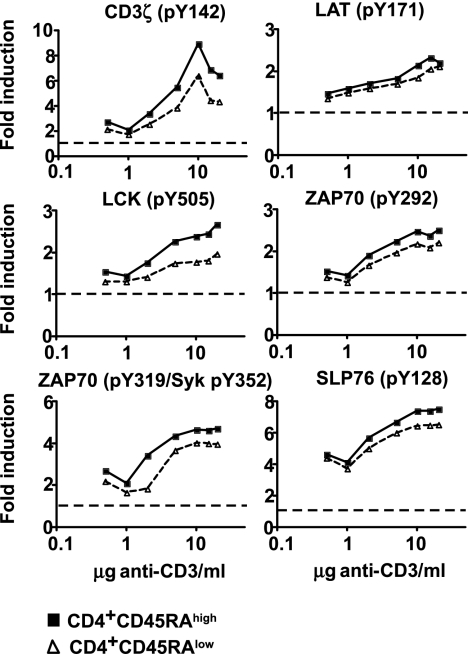
Fig. 2.
Induction of phosphorylation over a range of anti-CD3 concentrations. Phosphorylation was induced by incubation of whole blood at 37°C for 5 min. Titration results for both CD4+CD45RAhigh (filled squares) and CD4+CD45RAlow (open triangles) T cells are shown. Baseline levels are indicated by the dotted line. Donor-specific baseline levels are taken into account in the fold induction of phosphorylation.

