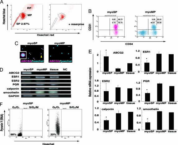
Fig. 1.
Isolation and characterization of human myoSP. (A Left) Distribution of the SP, MP, and replication (RP) populations of Hoechst 33342-stained living cells isolated from human myometrium. (Right) Coaddition of 50 μM reserpine resulted in the disappearance of the myoSP fraction. (B) CD31 and CD34 expression in myoSP and myoMP. (C) ABCG2 expression in myoSP and myoMP as determined by immunocytochemistry. Arrowheads indicate ABCG2-positive cells, one of which, indicated by a yellow arrowhead, is magnified (Inset). (Scale bar, 10 μm.) (D) mRNA expression of ABCG2, ovarian steroid receptors and smooth muscle cell markers in myoSP, myoMP, and whole myometrial tissues as determined by RT-PCR. NC, negative control (no RNA samples). (E) Relative mRNA expression of the ovarian steroid receptors and smooth cell markers in myoSP, myoMP, and whole myometrial tissues was examined by RT-PCR and normalized for GAPDH expression. Each bar indicates the mean + SEM of the relative expression obtained from three independent experiments using three individual samples. *, P < 0.05, versus myoMP. (F) Cell cycle status of myoSP and myoMP was determined by Hoechst 33342 and Pyronin Y staining. The left lower quadrant corresponds to the G0 phase.

