Fig. 3.
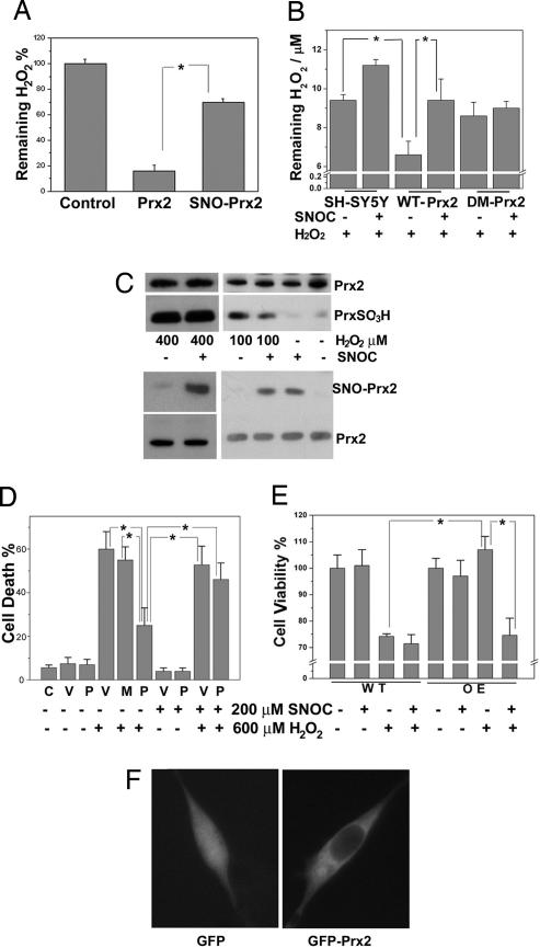
S-nitrosylation of Prx2 impairs its antioxidant function. (A) Chemical reduction of H2O2 by Prx2 or SNO-Prx2 in vitro. Recombinant Prx2 or SNO-Prx2 (after dissolution of any remaining free NO) was incubated with H2O2 for 20 min at room temperature. The remaining H2O2 level was assayed by using an H2O2 detection kit. *, P < 0.01 (n = 3). (B) SNOC inhibits reduction of H2O2 by Prx2 in SH-SY5Y cells. SH-SY5Y cells or SH-SY5Y cells stably overexpressing WT-Prx2 or double-mutant Prx2(C51A/C172A) (DM-Prx2) were pretreated with 200 μM SNOC before H2O2 challenge. Ten minutes later, the remaining H2O2 level was assayed. *, P < 0.01 (n = 4). (C) S-nitrosylation of Prx2 prevents its overoxidation by 100 μM H2O2. SH-SY5Y cells were exposed to 200 μM SNOC or control for 30 min and then incubated in 100 or 400 μM H2O2 for 10 min. Overoxidized forms of Prx, PrxSO2/3H, were detected by Western blotting using an antibody specific for PrxSO2/3H, and SNO-Prx2 was detected by the biotin-switch assay. (D) S-nitrosylation of Prx2 inhibits its protective function against H2O2-induced cytotoxicity in transiently transfected cells. SH-SY5Y cells were transiently transfected with WT Myc-Prx2 or C51A/C172A mutant Myc-Prx2. Cells were preexposed to SNOC or control for 30 min and then incubated in H2O2 for 24 h. Cell death was assayed by PI and Hoechst staining by counting the ratio of PI-positive (dead) to Hoechst-positive (total) cells. C, control; V, pCMV vector; P, myc-Prx2; M, C51A/C172A myc-Prx2. *, P < 0.05 (n = 4). (E) S-nitrosylation of Prx2 inhibits its protective function against H2O2-induced cytotoxicity in stably transfected cells. WT SH-SY5Y cells (WT) or SH-SY5Y cells stably overexpressing myc-Prx2 (OE) were preexposed to SNOC or control for 30 min and then incubated in H2O2 for 24 h. Cell viability was assayed by fluorescence intensity based on the metabolism of fluorescein diacetate to fluorescein in living cells. *, P < 0.05 (n = 4). (F) Overexpression of Prx2 does not affect its intracellular localization. SH-SY5Y cells were transiently transfected with GFP or GFP-Prx2. Prx2 distribution was monitored by deconvolution microscopy 24 h later.

