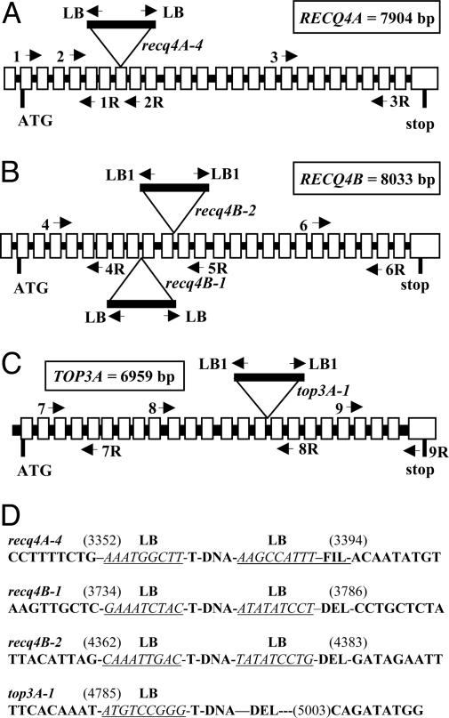
Fig. 1.
Molecular analysis of the T-DNA insertion lines. (A–C) The respective location of the T-DNA insertion in lines recq4A-4, recq4B-1 and 2, and top3α-1 is depicted. The schematically drawn genes of RECQ4A and RECQ4B contain 25 exons (gray boxes) and 24 introns (black lines) in the coding region and one additional exon and intron in the 5′ UTR, each. Both genes are interrupted in their helicase domains by the respective T-DNAs. The gene of TOP3α (At5g63920) contains 24 exons vs. 23 introns and the T-DNA is inserted in the 15th intron. Primers used are indicated by arrows. (D) Border sequence analysis of the insertion loci. The genomic sequences adjacent to each T-DNA insertion locus were determined by PCR and are depicted. In the case of top3α-1 on the right side, a deletion of ≈200 bp occurred. The genomic sequences are shown in bold, the respective left border sequence is in italics and underlined. FIL, filler sequence; DEL, deletion.

