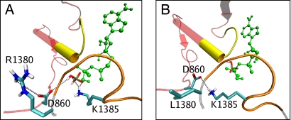
Fig. 6.
Wild-type and mutant ATP-binding site 2. (A) Homology model of ATP-binding site 2 of wild-type SUR1, indicating the main interactions of the posthydrolytic inorganic phosphate (CPK colors). MgADP is green, the NBD1 backbone is red, the signature sequence of NBD1 is yellow, the NBD2 backbone is gray, and the Walker A domain of NBD2 is orange. (B) Mutant model, with the posthydrolytic phosphate unbound. Unlike the wild-type simulation, K1385 hydrogen bonds to D860.

