Fig. 5.
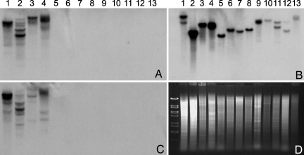
DNA blots of different species of Amanita. (A) Probed with AMA1 cDNA. (B) Probed with PHA1 cDNA. (C) Probed with a fragment of the β-tubulin gene isolated from A. bisporigera (see SI Text). (D) Ethidium-stained gel showing relative lane loading. Markers are λ phage DNA cut with BstEII. Species and provenances are as follows: lane 1, A. aff. suballiacea (Ingham County, MI); lane 2, A. bisporigera (Ingham County); lane 3, A. phalloides (Alameda County, CA); lane 4, A. ocreata (Sonoma County, CA); lane 5, A. novinupta (Sonoma County); lane 6, A. franchetii (Mendocino County, CA); lane 7, A. porphyria (Sonoma County); lane 8, a second isolate of A. franchetii (Sonoma County); lane 9, A. muscaria (Monterey County, CA); lane 10, A. gemmata (Mendocino County); lane 11, A. hemibapha (Mendocino County); lane 12, A. velosa (Napa County, CA); and lane 13, Amanital section Vaginatae (Mendocino County). Mushrooms represent sections Phalloideae (1–4), Validae (5–8), Amanita (9 and 10), Caesareae (11), and Vaginatae (12 and 13). Four separate gels were run; the lanes are in the same order on each gel, and approximately the same amount of DNA was loaded per lane. A and B are to the same scale, and C and D are to the same scale.

