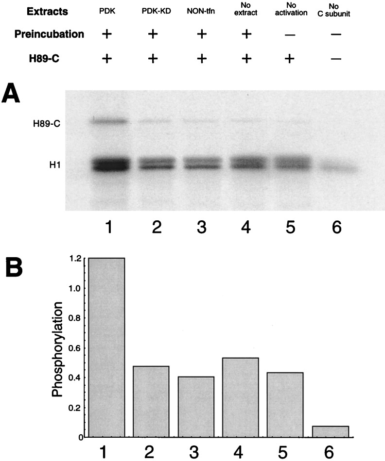
Figure 6.
Activation of PKA by PDK1. The S100 fractions of 293 cells (2.5 μl) independently transfected with Myc-PDK1 and Myc-PDK1-KD, as well as nontransfected 293 cells were immunoprecipitated by Myc antibodies and protein G-Sepharose as in Fig. 1. The immunoprecipitates were then mixed with histidine-tagged (H89)-C (0.125 μg) in the presence of [γ-32P]ATP. After incubation at room temperature for 30 min, the protein G beads were removed by centrifugation and H1 (1 μg) was added. The reaction mixtures were further incubated at room temperature for additional 10 min. Phosphorylated H1 was resolved by SDS/PAGE and visualized by autoradiography. Phosphorylation of H1 was quantitated by phosphoimaging. Units are arbitrary.

