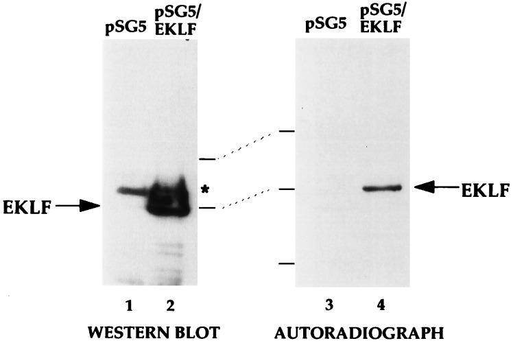
Figure 3.
Status of EKLF acetylation in vivo. COS7 cells transfected with 10 μg of pSG5 or pSG5/EKLF as indicated were labeled with sodium [3H]acetate in the presence of TSA and extracts were immunoprecipitated with anti-EKLF monoclonal antibody 6B3. Immunoprecipitated samples were resolved on SDS/PAGE and blotted and probed with anti-EKLF (Left) or processed and exposed in autoradiography (Right). Asterisk indicates a nonspecific signal from the immunoprecipitating antibody. Locations of molecular mass markers (70, 50, and 33 kDa, top to bottom) and EKLF are shown.

