Figure 3.
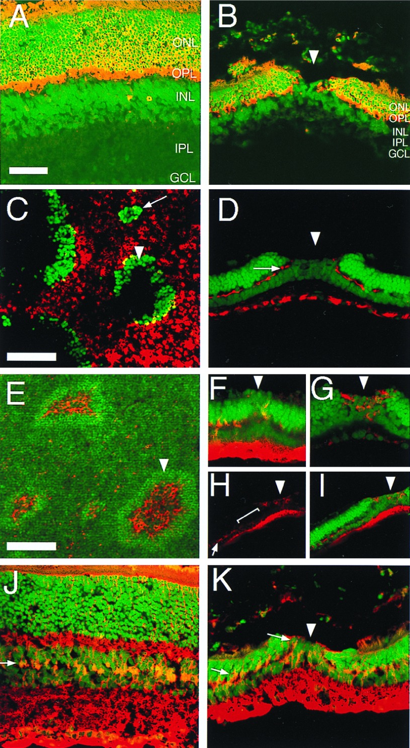
Immunohistochemical characterization of holes in the cyclin D1 mutant retinas. (A and J) Sections of P21 wild-type retinas. (B, D, F–I, and K) Sections of P21 cyclin D1−/− retinas. (C and E) Whole mount of P21 cyclin D1−/− retinas. Red, signals of antibody staining; green, nuclei. Arrowheads point to examples of holes. (A and B) Anti-recoverin; note the absence of recoverin-positive cells in the hole B. (C) Anti-rhodopsin, showing the outer segment surface of a mutant retina; note the absence of rhodopsin inside the holes. The arrow points to a small cell cluster that protrudes into the outer segment layer. (D) Anti-neurofilament; note the absence of neurofilament-positive cells in the hole. The arrow points to the neurofilament-positive horizontal cells outside the hole. (E–I) VC1.1 antibody; note the high density of VC1.1-positive cells inside the holes and reduction of VC1.1-positive cells adjacent to the holes. (E) A whole mount retina stained with VC1.1 antibody. (F–I) VC1.1 antibody staining of sections. (F) A hole at early stage. (G) A hole at late stage. (H) VC1.1 antibody staining of a hole at low magnification. Arrow in H points to the VC1.1-positive cells at a distance to the hole. Bracket in H indicates the area that contains fewer VC1.1-positive cells. (I) Merged image of the VC1.1 antibody staining in H and the nuclear stain. (J and K), anti-CRALBP; note the location of the CRALBP-positive Müller glial cells in the middle of the INL in the wild-type (arrow in J) and the abnormal location of the Müller glial cells in the ONL in the cyclin D1−/− mutant (arrows in K). OPL, outer plexiform layer. IPL, inner plexiform layer. GCL, ganglion cell layer. (Scale bar in A is 50 μm; A, B, J, and K are to the same scale. Scale bar in C is 50 μm; C, D, F, and G are to the same scale. Scale bar in E is 25 μm; E, H, and I are to the same scale.)

