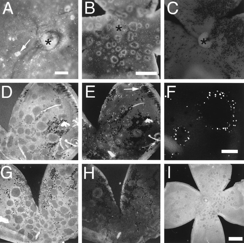
Figure 4.
Apoptosis in the cyclin D1−/− mutant retinas. In all panels, the photoreceptor side of the whole mount retina is shown (vitreous side facing down). (A–C) Acridine orange staining. (A) Cyclin D1−/−, P6. Arrow points to a cluster of acridine orange-positive cells. (B) Cyclin D1−/−, P10; note the rings of acridine orange-positive cells and some scattered acridine orange-positive cells. (C) Wild-type. Asterisks indicate the optic nerve head. (D–F) TUNEL assays of P21 cyclin D1−/− mutant retinas. (D) Dark field image of the retina. (E) TUNEL assay viewed under UV illumination; note the presence of TUNEL-positive cells at the edge of the holes. One of the holes is indicated by the arrow and is shown at a higher magnification in F. (G and H) Negative control for TUNEL assay (without adding terminal transferase in the assay). (G) Dark field image. (H) UV illumination. (I) P10 cyclin D1−/−; opsin/bcl-2. Note the presence of holes. Black speckles in retinas were cells or pigment granules from the pigmented epithelium that adhered to the retinas. (Scale bar in A is 100 μm. Scale bar in B is 250 μm; B, C, D, E, G, and H are to the same scale. Scale bar in F is 50 μm. Scale bar in I is 500 μm.)

