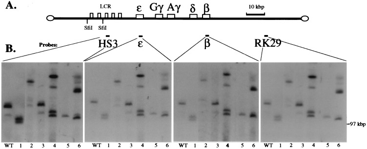
Figure 2.
Mapping the integrated Aγ3′E YAC transgene structures. (A) Diagram of the human β-globin YAC showing the SfiI sites and probes used. (B) Analysis of the integrity of the YAC transgenic lines. High molecular mass DNA isolated from thymus cells of transgenic lines was digested with SfiI and subjected to PFGE (see Materials and Methods). The gel was blotted to nylon filters and hybridized to probes corresponding to HS3, ɛ- and β-globin genes, and 3′ flanking marker RK29, respectively, in the four panels shown. The position of the bacteriophage λ dimer marker run in parallel (97 kbp) is shown on the right. All four probes hybridize to SfiI fragments larger than 100 kbp, and each probe hybridizes to the identical bands in each transgenic line.

