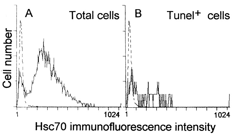
Figure 6.
Flow cytometric analysis of Hsc70- and TUNEL-stained cells from E1.5 embryos cultured for 6 h in basal medium. Double TUNEL and Hsc70 immunostaining was performed in dissociated cells. (A) One representative Hsc70 staining (continuous line) of total cells (of 15 embryos in five independent experiments). Nonimmune Ig was used as a control (dotted line). (B) Hsc70 immunofluorescence levels were also analyzed in the TUNEL-stained cells. Note that 45% of apoptotic cells coincided with the area considered negative for Hsc70 (dotted line) and 90% of the TUNEL-stained cells showed an Hsc70 immunofluorescence signal below the mean signal of the total cell population. The profiles were similar for embryos cultured with insulin, but the proportion of apoptotic cells was lower.

