Figure 2.
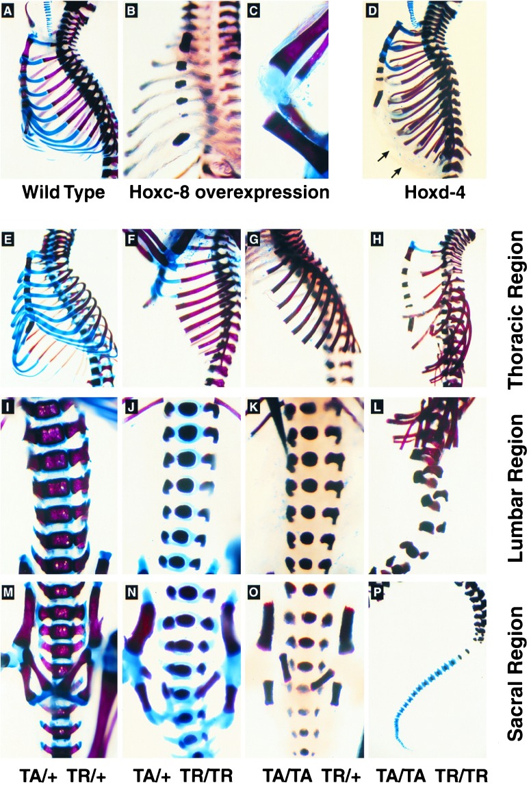
Cartilage abnormalities in Hox gene transgenic mice. Skeletons from newborn mice were stained with Alizarin red (bone) and Alcian blue (cartilage). (A–D) Cartilage defects upon overexpression of Hoxc-8 (B and C) or Hoxd-4 (D) transgenes. (A) Skeleton of a wild-type newborn FVB mouse. (B and C) Reduced Alcian blue staining in ribs and knee cartilage of a Hoxc-8 transgenic mouse that died shortly after birth. (D) Alcian blue staining was reduced in ribs and vertebral column of a Hoxd-4 transgenic animal. Note that the cartilaginous portions of the ribs were present (arrows in D, compare with B) and that tracheal cartilage was normal. (E–P) Severity of cartilage abnormalities increased with transgene dosage. The skeleton from an animal hemizygous for both TA and TR loci resembled staining of the wild-type situation (compare E to A). The thoracic (E–H), lumbar (I–L), and sacral regions (M–P) of the skeletons from animals with the genotypes TA/+ TR/+ (E, I, M), TA/+ TR/TR (F, J, N), TA/TA TR/+ (G, K, O), and TA/TA TR/TR (H, L, P) are shown. All animals with transgene loci in excess of hemizygosity died shortly after birth or were dead at birth. The distortion and flexibility of the skeleton were highest in the animal homozygous for both transgenes.

