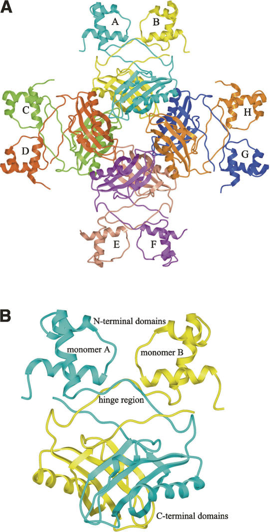
Figure 3.
(A) Schematic representation of the M. tuberculosis LrpA octamer, showing the dimer–dimer interactions that lead to the formation of a stable octamer. Each subunit is labeled A–H, and colored as follows: A, cyan; B, yellow; C, light green; D, rust; E, light salmon; F, purple; G, blue; H, orange. (B) Schematic representation of the M. tuberculosis LrpA dimer, with local twofold noncrystallographic symmetry, demonstrating the LrpA subunit–subunit interactions that promote the formation of a stable dimer. Subunit A is colored in cyan and subunit B is colored in yellow.

