
Figure 2.
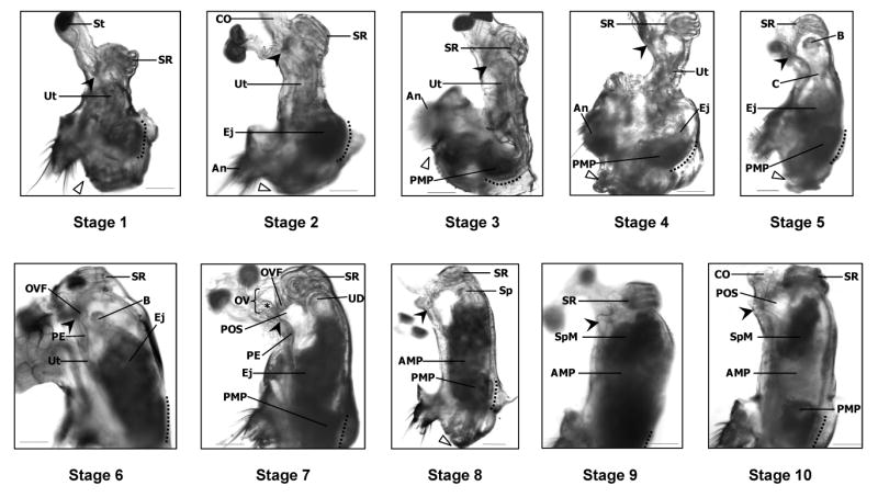
Stages of sperm transport in D. melanogaster. In virgin (Oregon R) females (Stage 1), the uterus (Ut) is highly compacted and the seminal receptacle (SR) is pulled ventrally. Soon after copulation begins (Stage 2), the uterus lengthens and ejaculate (Ej) begins to accumulate at the specialized vaginal intima (Miller 1950; SVI, to the left of dotted line). As ejaculate transfer continues (Stages 3–6), the uterus unfolds and takes on a turgid, oval shape. By Stage 7, the oviduct valve flap (OVF) has moved anteriorly, forming the top of the pre-oviduct space (POS). In Stage 8, sperm (Sp) begin to move toward the anterior uterus, leaving behind the lightly shaded anterior mating plug (AMP), visible in mid-uterus, and the posterior mating plug (PMP) adjacent to the SVI. Sperm accumulate in a mass (SpM) at the anterior uterus (Stage 9) where the openings to the sperm storage organs are located; these organs are out-of-focus in this picture. Sperm are also visible within the seminal receptacle. Finally, in Stage 10, the size of the SpM has decreased, presumably reflecting the movement of sperm into storage. The location of sperm in each stage was corroborated by examining the reproductive tracts of Oregon R females that had mated with dj-GFP (Santel et al. 1997) males, whose sperm are GFP-labeled. Filled arrowheads indicate the point where the spermathecal ducts open into the uterus. Open triangles mark the gonopod long bristle, located near the dorsal edge of the vulva. Dotted lines parallel the SVI. An, anus; B, “ball” of sperm (see text for details); C, “column” of sperm (see text for details); CO, common oviduct; St, spermatheca; OV, oviduct valve; PE, papillate elevation (Miller 1950); UD, uterus dome; *, ridge in dorsal oviduct wall which makes up part of the OV (Scale = 0.01mm).