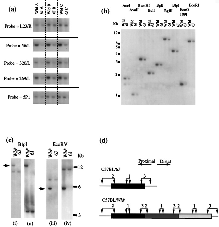
Figure 2.
Identification by genomic Southern blotting of a tandemly repeated region on Wlds chromosome 4. (a) Three probes (56/L, 320/L, and 269/L) lying within 30 kb of one another show an amplified signal when hybridized to a Southern blot of Wld genomic DNA. Markers on either side (L23/R and 5P1) show an approximately equal signal in C57BL/Wlds and C57BL/6J when hybridized to the same filter. (b) Probes located centrally within the repeat unit (320/L is shown here) consistently show an increase in signal intensity but no difference in restriction fragment size with a range of frequent-cutter restriction enzymes. (c) Probe 59 (i) and (iii) located within the repeat unit immediately adjacent to the proximal boundary, hybridizes to a fragment of identical size in C57BL/Wlds and in C57BL/6J, but also to a Wld-specific fragment of altered size and with an increased intensity. A probe located 0.8–1.0 kb from the distal end of the repeat unit (ii and iv) detects the same Wld-specific fragment (indicated by arrows) as probe 59, together with a different fragment that is common to both C57BL/6J and C57BL/Wlds. (d) Tandem repeat model to account for the above observations. Probe 1 represents probes used in a and b. It lies centrally in the repeated region and detects a restriction fragment of increased dosage but unaltered size in C57BL/Wlds. Probe 2 represents probe 59 in c. It detects one restriction fragment that is present as a single copy in both C57BL/6J and C57BL/Wlds. However, when the target sequence of this probe is repeated in C57BL/Wlds, a shifted band appears because of the formation of a junction fragment. This band may be shifted up or down depending on the location of the restriction site shown on the left. Probe 3 is located at the other end of the repeat unit and detects the same shifted fragment as probe 2, but a different fragment that is common to both C57BL/6J and C57BL/Wlds.

