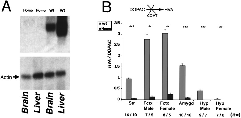
Figure 2.
(A) Northern blot analysis of mRNA isolated from the liver and the brain of homozygous and wild-type animals. Two mRNA species are observed in the liver, corresponding (by analogy to the rat and human gene) to two distinct sites of transcriptional initiation. The coding part of the COMT cDNA was used as probe. As a control the Northern blot was probed with a probe from rat β actin. (B) HVA/DOPAC ratio in the striatum, frontal cortex, amygdala, and hypothalamus of male and female homozygous and wild-type animals. Values are the average ± SEM for wild-type (shaded bar) and homozygous (solid bar) mice. Differences between wild-type and homozygous mice tested by Mann–Whitney u test were ∗∗∗, P < 0.0002, ∗∗, P < 0.002. The calculated HVA/DOPAC ratios for homozygous (Homo) versus wild-type (wt) animals are as follows: HVA/DOPACstriatum = 0.05 ± 0.009 vs. 0.94 ± 0.71 (P = 0.0001); HVA/DOPACfrontal cortex/males = 0.13 ± 0.04 vs. 2.77 ± 0.59 (P = 0.003); HVA/DOPACfrontal cortex/females = 0.25 ± 0.09 vs. 3.05 ± 0.37 (P = 0.002); HVA/DOPACamygdala = 0.09 ± 0.009 vs. 1.56 ± 0.17 (P = 0.0002); HVA/DOPAChypothalamus/males = 0 vs. 0.41 ± 0.05 (P = 0.0006); and HVA/DOPAChypothalamus/females = 0 vs. 0.04 ± 0.008 (P = 0.002); It is notable that although the HVA/DOPAC ratio is decreased significantly in all of the brain areas tested, residual HVA is detectable, in several of these areas (not shown). One interpretation is that the residual methylation of dopamine and DOPAC is caused by the action of yet unidentified methyltransferases involved in the clearance of dopamine, whose activity is probably up-regulated in the absence of COMT. Further work is needed to verify this interpretation and possibly lead to better characterization of this pathway. Str, striatum; Fctx, frontal cortex; amygd, amygdala; Hyp, hypothalamus. The number of the animals tested (n =) is indicated.

