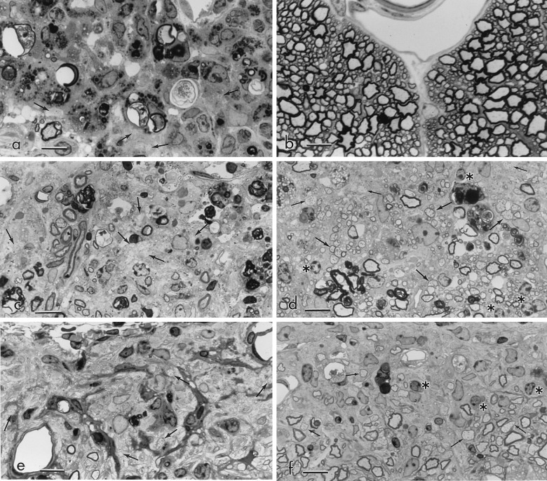
Figure 2.
Histopathology of spinal cord vehicle- and rhGGF2-treated mice; 1 μm epoxy sections; toluidine blue stain. (Bar = 10 μm.) (a) Acute EAE. Experiment 3: Vehicle treatment 1–10 dpt; sampled 11 dpt; clinical grade 4. An area of subpial spinal cord displays extensive inflammation. Note the many myelin-laden macrophages and scattered demyelinated axons (arrows). (×875.) (b) Acute EAE. Matching level of spinal cord to a, treated with rhGGF2 (2 mg/kg) 1–10 dpt; sampled 11 dpt; clinical grade 0.5. A matching area of spinal cord displays no lesion activity. Oligodendrocytes difficult to discern. (×875.) (c) Chronic relapsing EAE. Experiment 4: Vehicle 31–55 dpt; sampled 60 dpt; clinical grade 3. A chronic gliotic lesion contains macrophages and displays many demyelinated axons (arrows). Oligodendrocytes difficult to discern. (×875.) (d) Chronic EAE. rhGGF2 (2.0 mg/kg) 31–55 dpt; sampled 60 dpt; clinical grade 3.5; spinal cord. Despite the comparable clinical grade of this matching animal to that shown in (c), note the large amount of CNS remyelination (large arrows). Some demyelinated axons (small arrows) lie toward the subpial surface. Numerous oligodendrocytes (∗) identified by their rounded nuclei and clumped heterochromatin, can be seen. (×875.) (e) Chronic relapsing EAE. Experiment 7: Vehicle 21–79 dpt; sampled day 81; clinical grade 3. A subpial lesion from the L7 spinal cord level displays intense fibrous astrogliosis, fibrotic blood vessels, and numerous demyelinated axons (arrows), but no obvious oligodendrocytes. (×875.) (f) Chronic relapsing EAE. rhGGF2 (0.2 mg/kg and 0.02 mg/kg) days 21–79; sampled day 81; clinical grade 2. Matching animal to e, same level of spinal cord. Note the more compact, less gliotic parenchyma and presence of remyelinated CNS fibers (arrows). A few oligodendrocytes (∗) are present. (×875.)

