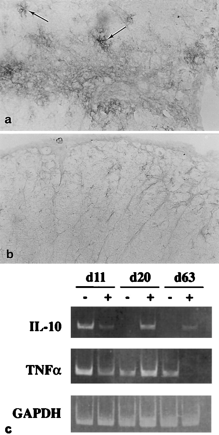
Figure 4.

Th2-type cytokine expression and GGF2-treatment. (a) Lumbar spinal cord; rhGGF2-mouse (1.0 mg/kg; 9–40 dpt), sampled 42 dpt shows high level immunoreactivity for IL-10 on perivascular and parenchymal astrocytes (arrows) bordering a penetrating blood vessel. (×250.) (b) A comparable area of lumbar spinal cord from a control mouse at 42 dpt displays low level reactivity for IL-10. (×250.) (c) reverse transcription–PCR analysis of CNS tissue. Representative mice (n = 6) from acute (11 dpt), remission (20 dpt) and chronic (63 dpt) phases of EAE, from vehicle (−) and rhGGF2-treated (+) groups for analysis of cytokine gene expression. At 11 and 20 dpt, the rhGGF2-treated animals were from the 2.0 mg/kg group, whereas at 63 dpt, animals from the 0.8 mg/kg group were selected. RNA from spinal cord was analyzed by reverse transcription–PCR using specific primers for IL-10, TNFα, or glyceraldehyde-3-phosphate dehydrogenase. DNA amplification products analyzed by gel electrophoresis were 455 bp, 354 bp, and 356 bp, respectively. Note increased levels of IL-10 RNA in rhGGF2-treated animals at days 20 and 63 dpt, and the marked decrease of TNFα at 63 dpt in the same group.
