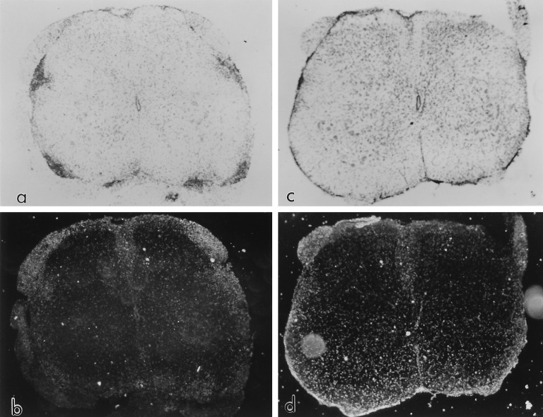
Figure 5.
In situ hybridization for MBP exon 2 message.(a) Bright field microscopy shows punctate lesion activity in a vehicle-treated mouse sensitized for EAE. Vehicle; 1–40 dpt; sampled 64 dpt; clinical grade 2.5. No counterstain. (×20.) (b) Same section as in a, in situ hybridization, developed for anti-sense MBP exon 2 message. Background levels of MBP exon 2 mRNA seen throughout the spinal cord with some elevation over dorsal roots (above). (×20.) (c) Matching animal to a treated with rhGGF2 (0.8 mg/kg) 0–40 dpt; sampled 64 dpt; clinical grade 3.5. No obvious lesions apparent other than some inflammation over the spinal cord. No counterstain (×20.) (d) Same section as in c, in situ hybridization, developed for MBP exon 2 message. Note enhanced signal for MBP exon 2 over anterior and lateral columns, probably indicative of diffuse CNS remyelination. Some elevation of signal occurs over the dorsal roots and meninges. (×20.)

