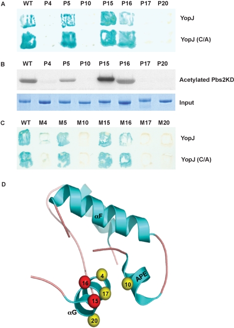
Figure 6. The binding site on MKKs for YopJ.
(A) Yeast two-hybrid assay of the interaction between YopJ or the YopJ(C172A) mutant and wild type Pbs2 or mutants of Pbs2 in or around G α-helix. (B) Recombinant YopJ is incubated with purified Pbs2 kinase domain or mutants in or around the G α-helix in the presence of 14C-labeled acetyl-CoA for 1 hour at 30°C. Samples are separated by SDS-PAGE and analyzed by autoradiography. Substrate loading was controlled by protein staining. (C) Yeast two-hybrid assay of the interaction between YopJ or YopJ (C172A) mutant and corresponding MKK1 mutants. (D) Structural mapping of G α-helix and APE domain, showing the mutants tested above.

