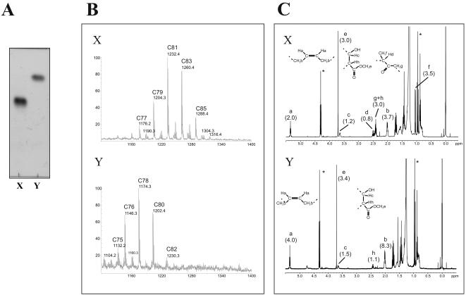
Figure 4. Structural determination of lipids X and Y.
Lipids X and Y were purified from cell wall extract of M. bovis BCG Pasteur culture, following treatment with SRI-224 (5 µg/ml) for 24 h. (A) Conventional TLC showing purity of the samples containing lipids X and Y, that were used for structural analyses, as seen by staining with phosphomolybdic acid and charring. (B) m/z values from MALDI-TOF-MS spectra correspond to [M+Na]+ adducts of a family of methylated keto-mycolates and α-mycolates for purified lipids X and Y, respectively. (C) For 1H-NMR analysis, protons are labelled (a to h) according to their respective positions in functional groups. Relative integrations of protons have been normalized according to the number of ethylenic protons (2 for X and 4 for Y) and are indicated in brackets. *stands for proton 1H signals of contaminant ethanol present in the NMR tubes.

