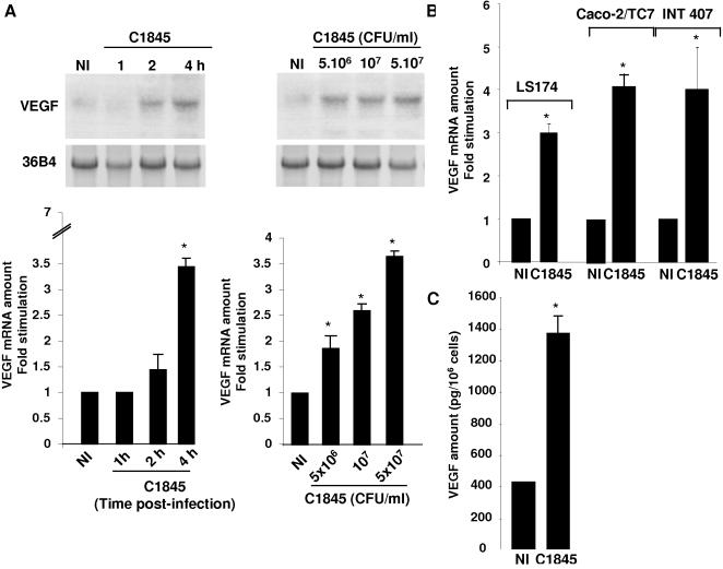
Figure 1. C1845 bacteria increase VEGF expression in cultured intestinal epithelial cells.
In A, increase in VEGF mRNA expression. Confluent serum-starved T84 cells (5×10⁶ cells/well) were infected with wild-type C1845 bacteria and total RNA was prepared as indicated in the Materials and Methods section. Results for non-infected (NI) or cells infected with 5×10⁷ CFU/ml C1845 bacteria for the indicated time are shown on the left panel. The dose response effect is presented on the right. Cells were infected for four hours with the indicated number of bacteria. Results of Northern blots are presented in the upper part of the Figure and q-PCR shown in the lower part. These results are representative of three independent experiments. In B, confluent serum-starved LS174, Caco-2/TC7 or INT407 cells were infected with 5×10⁷ CFU/ml wild-type C1845 bacteria for four hours. VEGF mRNA expression was assayed by q-PCR. The signal corresponding to VEGF and 36B4 transcripts was quantified using a phosphoImager. Under each condition the signal was normalized to the 36B4 probe. Results are expressed as arbitrary units corresponding to the fold stimulation of treated versus non-treated conditions. In C, increase in the VEGF protein in the culture medium of wild-type C1845-infected T84 cells. Cells were infected with 5×10⁷ CFU/ml wild-type C1845 bacteria for four hours and the supernatant were collected as indicated in the Materials and Methods section. The VEGF protein level was then quantified using an ELISA. Quantification of the results from two independent experiments (means±SD) is shown. *, p<0,01

