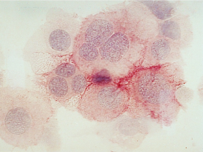
Figure 4.
Immunohistochemical staining of enriched H/RS cells (case 6937) on a cytospin slide. The nuclei of the cells are stained with hematoxyline and CD30 expression is visualized by avidin-biotin-complex staining. The giant H/RS cells are clearly visible. Some of the cells are classical RS cells with more than one nucleus. (×630.)

