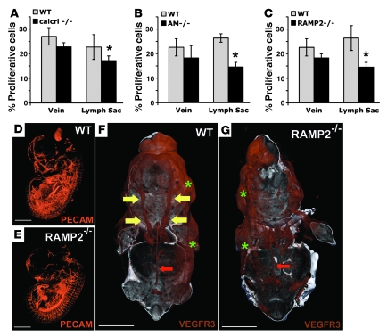
Figure 7. AM signaling is required for the proliferation and growth of jugular lymphatic vessels during embryonic development.
(A–C) The percentage of proliferating cells relative to total cells was determined in the jugular vein and neighboring jugular lymph sac of wild-type (gray bars) and calcrl–/–, AM–/–, and RAMP2–/– (black bars, A–C, respectively) littermate embryos. Percent proliferative cells was defined as the number of BrdU-positive endothelial cells divided by the total number of endothelial cells in each vessel. Error bars indicate SEM. *P = 0.01 for calcrl–/– (A), P = 0.004 for AM–/– (B), and P = 0.01 for RAMP2–/– (C); Student’s t test. n > 6 animals per genotype. (D and E) Whole mount immunofluorescence of developing vasculature identified by PECAM staining and visualized by 3D OPT of wild-type (D) and RAMP2–/– (E) littermate embryos at E13.5. (F and G) Whole mount immunofluorescence of developing lymphatic vasculature identified by VEGFR3 staining and visualized by 3D OPT of wild-type (F) and RAMP2–/– (G) littermate embryos at E14.5. Compare the presence of well-formed jugular lymph sacs in the wild-type embryo (yellow arrows in F) with the relative lack of jugular lymphatic vessels in the RAMP2–/– littermate (G). Note that the retroperitoneal lymph vessel (red arrows) and dermal lymphatic vessels (green asterisks) of RAMP2–/– mice appear normal compared with those of wild-type embryos. Three embryos from 2 different litters were stained for PECAM and VEGFR3. The online supplemental data contains movies for enhanced, high-resolution, 3D viewing (Supplemental Figures 4 and 5). Scale bar: 2,000 μM.

