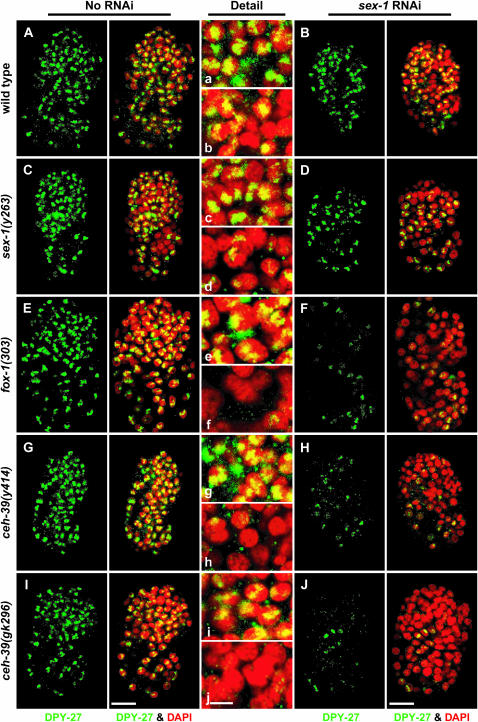
Figure 3.—
Decreasing the dose of ceh-39 and other XSEs disrupts the dosage compensation complex in XX animals. Localization of DPY-27 in wild-type and XSE mutant embryos with and without RNAi disruption of sex-1. (A–J) Partial projections of false-colored confocal images of wild-type and mutant XX embryos costained with antibodies against DPY-27 (green) and the DNA intercalating dye DAPI (red). (a–j) Enlargements of nuclei from A–J, respectively. (A, E, G, and I) DPY-27 localized in a punctate pattern to the X chromosomes of wild-type XX embryos, and fox-1 or ceh-39 mutant XX embryos. (C) The sex-1(y263) mutant embryos exhibited a reduction in DPY-27 staining. Some nuclei showed sparse or no staining and others varied from punctate to diffuse staining, all implying less DPY-27 on X. (B and D) sex-1(RNAi) XX embryos and sex-1(y263, RNAi) mutants showed a further decrease in DPY-27 signal compared to that in sex-1 mutants. The residual DPY-27 was mostly punctate, indicating X localization. (F, H, and J) The DPY-27 signal was drastically reduced in fox-1 sex-1(RNAi) XX and ceh-39 sex-1(RNAi) XX mutants; the small quantity of residual DPY-27 had punctate localization. The more severe reduction in DPY-27 signal after knockdown of two XSEs rather than one shows that dosage compensation is disrupted more in double than in single XSE mutants, as is viability. In the images, DPY-27 signal was enhanced in all embryos treated with RNAi of sex-1 to demonstrate the punctate localization more clearly. Bars: A–J, 10 μm; a–j, 3 μm.

