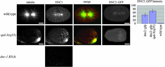
Figure 4.—
spd-3(oj35) embryos have defects in dynactin localization. In wild-type embryos, DNC-1 localizes to the centrosomes, the mitotic spindle, and the plus ends of astral microtubules (n = 12). oj35 embryos exhibit an additional, anomalous localization of DNC-1 to posterior structures resembling P granules (n = 12). DNC-1 signal is absent in dnc-1 RNAi embryos. DNC-2∷GFP in wild-type embryos is present throughout the cytoplasm, with enrichments in the pericentriolar region and along the mitotic spindle (n = 5). This enrichment is decreased in oj35 embryos, while the cytoplasmic signal is significantly increased (n = 6). DNC-2∷GFP images portrayed are snapshots taken from live imaging, while tubulin (n357) and DNC-1 antibodies were used with fixed embryos. Bar, 10 μm.

