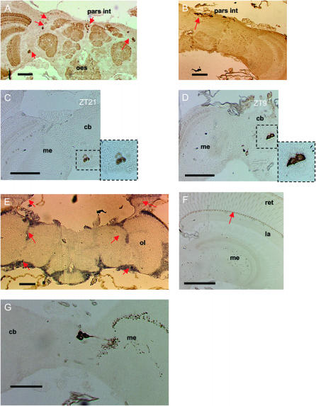
Figure 5.—
Clock protein expression in head sections of Musca. (A) A section at ZT21 labeled with αDmPER-I and represents a general staining pattern obtained with all available anti-PER antibodies. Several groups of cells are labeled (arrows), including the pars intercerebralis and a dorsal and ventral group of neurons that are lateral to the central brain. (B) Replacing the αDmPER-I antibody with rabbit normal serum results in staining of pars intercerebralis cells (arrow), suggesting that these are nonspecific for PER staining. (C and D) During both the night (ZT21) and the day (ZT9) anti-PER immunoreactivity was exclusively cytoplasmic with cells showing a characteristic “doughnut” shape. (E) In situ hybridization to Mdper. Arrows denote hybridization to regions where the dorsal and lateral PER-positive neurons are located and the photoreceptors. (F) Although no staining is observed in photoreceptor nuclei at night, a structure at the base of the photoreceptor can be seen to stain strongly at all times (arrow). (G) PDF-expressing cells and their projections. pars int, pars intercerebralis; oes, esophagus; me, medulla; la, lamina; ret, retina; ol, optic lobe. Bars, 100 μm.

