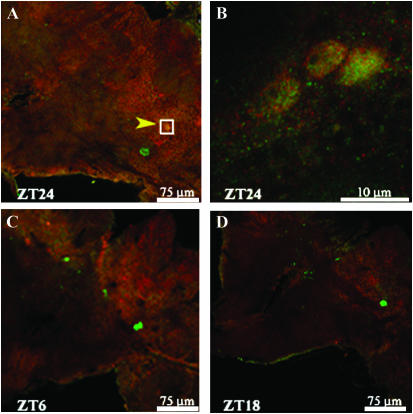
Figure 9.—
Confocal images of PER- and TIM-expressing neurons at different time points in Musca. (A) The PER-expressing medial lateral neurons are in the proximity of the PER- and TIM-expressing s-LNvs, here shown arrowed and boxed. (B) Larger magnification of the boxed region of A showing nuclear colocalization of PER and TIM in sLNvs at ZT24. (C and D) At ZT6 and -18, the same region shows the brightly stained medial lateral neurons but indicates the absence of PER and TIM staining in the sLNvs.

