Figure 4.—
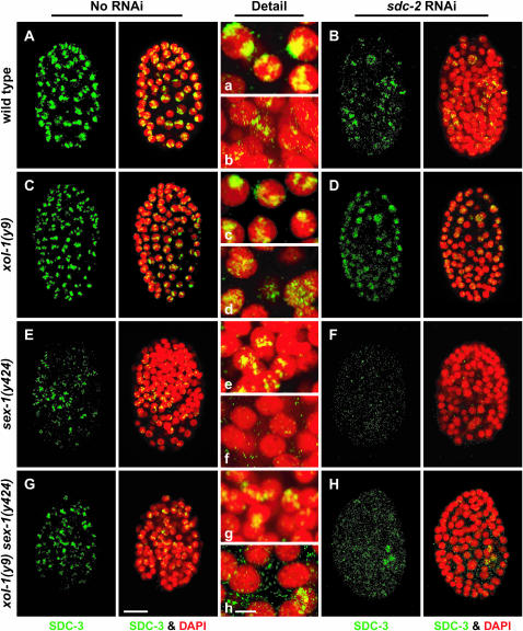
DCC disruption by a sex-1 null mutation and by the XSE-independent function of sex-1. (A–H) Partial projections of false-colored confocal images of wild-type and mutant XX embryos costained with antibodies against the dosage compensation protein SDC-3 (green) and DAPI (red). (a–h) Enlargements of nuclei from A–H, respectively. (A–D) RNAi disruption of sdc-2 in wild-type and xol-1 mutant embryos reduced the abundance and partially disrupted the X localization of SDC-3. (E and F) The sex-1(y424) null allele partially disrupted dosage compensation, but X-localized SDC-3 was evident in many nuclei. Some nuclei lacked SDC-3 staining. RNAi of sdc-2 into sex-1(y424) completely disrupted the DCC. Virtually no SDC-3 was detectable. (G and H) RNAi of sdc-2 in xol-1 sex-1 mutants also disrupted SDC-3 severely, similarly to xol-1(y9) sex-1(y424) sdc-2(RNAi) embryos, showing that the XSE-independent function of sex-1 is important for proper stability and assembly of the DCC on X. Bars: A–H, 10 μm; a–h, 3 μm.

