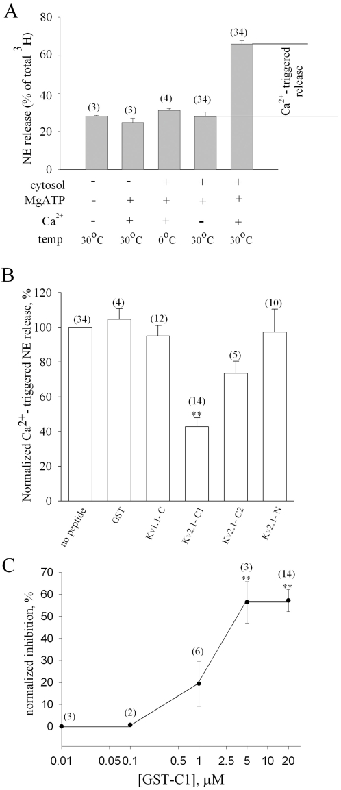
Figure 2. Kv2.1 syntaxin-binding peptides inhibit NE secretion.
(A) PC12 cells preloaded with [3H]-NE and gently homogenized ( “cracked” cells), either underwent 15 min incubation in the presence of 2 mM MgATP and brain cytosol at 30°C (“priming”) or not, followed by either 15 min incubation with added 1.6 mM Ca2+ (1 µM free Ca2+ ;[39] at 30° (“triggering”) or not, as indicated below the bars. Cells were pelleted and the secreted [3H]-NE in the supernatant was quantified by scintillation counting and expressed as a percentage of the total [3H]-NE in the cell. 106 cells per reaction were used. The difference between the amounts of [3H]-NE secreted in the two reactions on the two bars on the right was defined and is referred to hereafter as the Ca2+-triggered release. (B) Cracked cells preloaded with [3H]-NE underwent priming and triggering reactions in the absence or presence of 10–20 µM GST-fusion peptides corresponding to cytosolic parts of Kv2.1 and Kv1.1 (shown in Fig. 1A) or to GST itself (as indicated below bars). Values of Ca2+-triggered release measured in the presence of the peptides (see Fig. 2A) were normalized to the control release determined in the absence of peptides (defined as 100%). Each bar in A and B depicts the mean±s.e.m from several independent experiments (numbers of experiments are in parentheses above bars). **, p<0.001 (compared with GST). (C) Cells were stimulated to release NE in the presence of increasing concentrations of GST-fused Kv2.1-C1 protein. Each point in the curve represents mean±s.e.m values from several independent experiments (numbers of experiments in parentheses above bars). **, p<0.001 (compared with 10 nM GST-C1).

