
Figure 4.
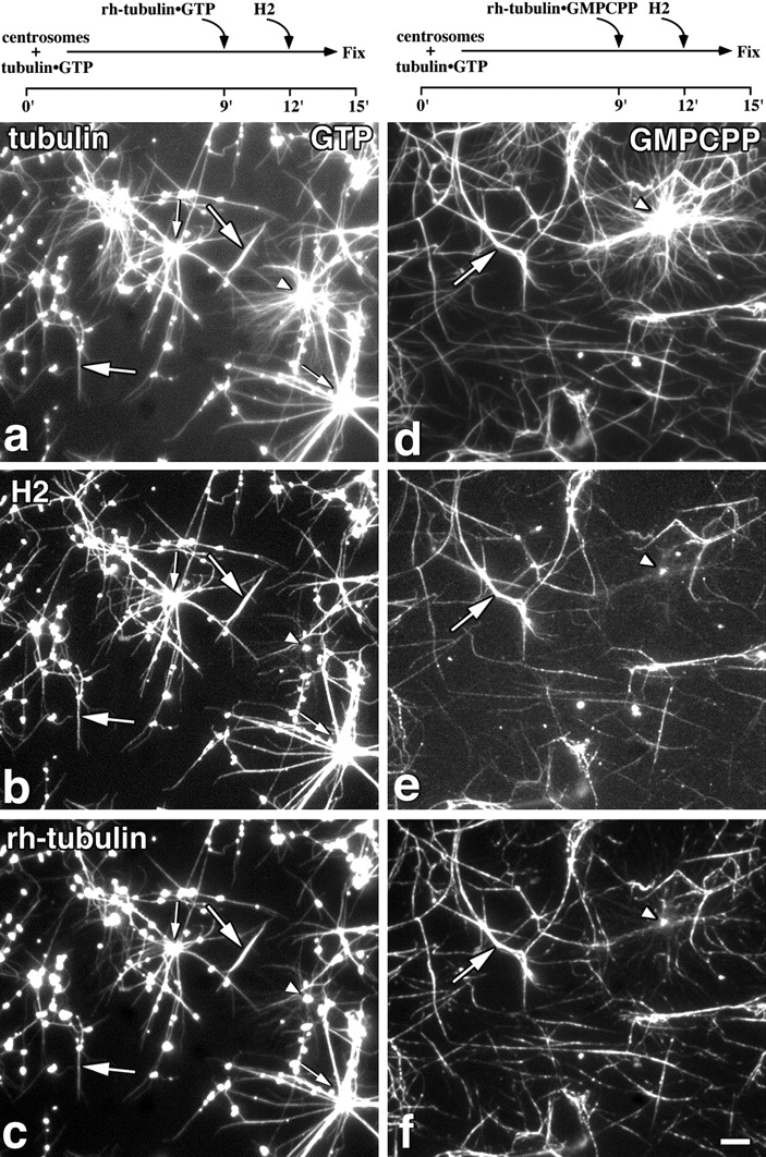
CLIP-170 binds preferentially to newly polymerized microtubules in vitro. Tubulin (∼1 mg/ml) was polymerized from centrosomes in the presence of 1 mM GTP for 9 min before a second addition of rh-tubulin, at 0.3 mg/ml equilibrated with GTP (a–c) or at 0.1 mg/ml equilibrated with GMPCPP (d–f). The final nucleotide concentrations were 1.2 mM GTP for a–c and 1.0 mM GTP, 0.1 mM GMPCPP for d–f. After a further 3 min of incubation, H2 was added at 40 μg/ml and the asters were fixed 3 min later (experimental time line above relevant panels). The fixed microtubules were sedimented onto coverslips as described in Materials and Methods and labeled for tubulin (a and d) using a monoclonal antibody (1A2) or H2 (b and e) using an anti-peptide antibody (anti-KRKV). Rh-tubulin is shown in c and f. Arrowheads, asters that have not incorporated rh-tubulin, and are not labeled for H2; small and large arrows, rh-labeled asters and rh-labeled free microtubules, respectively, which are also labeled for H2. Bar, 10 μm.