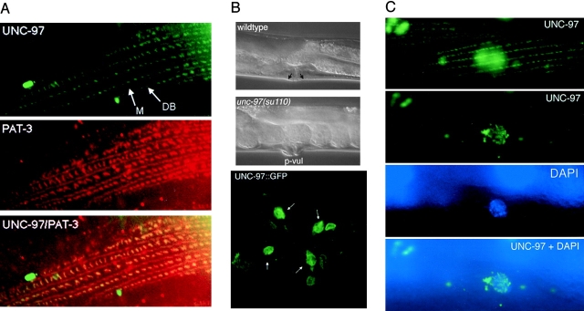
Figure 6.
Subcellular localization of UNC-97. (A) Top, UNC-97::GFP localization to dense bodies (DB) and M lines; middle, PAT-3 localization visualized with the anti-MH25 antibody; bottom, overlay of the first and second panel showing the overlap between green (UNC-97::GFP) and red (PAT-3) in yellow. (B) Top two panels show lateral Nomarski images of wild-type and unc-97(su110) animals, the latter animal displaying a protruding vulva phenotype (p-vul). The p-vul phenotype displays a 56% penetrance (n = 32). Black arrows in top panel, approximate attachment sites of the vulval muscles. The shape of the vulval muscles can also be seen in Fig. 4 D. Bottom panel, ventral view of UNC-97::GFP localization in wild-type adult animals. UNC-97::GFP localizes to the attachment sites (white arrows) of the vulval muscles to the hypodermis (for a schematic drawing of the vulval muscles see White, 1988). (C) Top, UNC-97:: GFP localization in the muscle sarcomere. Thin stripes, M line structures; dots, dense body structures (for a schematic description of these structures see Fig. 1). In the center of the panel, fuzzy nuclear staining can be observed which is out of focus. Second panel, the same cell as in the first panel, but in a different plane of focus showing nuclear localization of UNC-97 in dots. Third panel, DAPI staining of the same nucleus. Bottom, overlay between the second and third panel. The animal shown in this series of micrographs has been fixed with formaldehyde to stabilize sarcomeres and nuclear localization of UNC-97.

