
Figure 1.
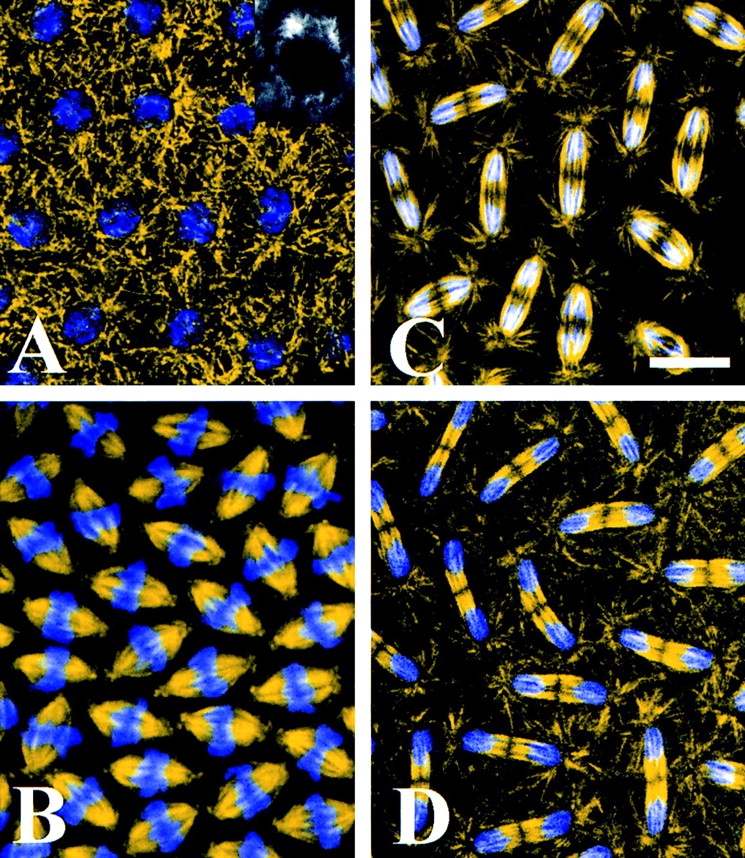
Mitosis in the syncytial blastoderm of early Drosophila embryos. Confocal immunofluorescence images of embryos immunostained with an antibody raised against Drosophila embryonic tubulin (yellow) overlaid with the corresponding DNA stain DAPI (blue). (A) Prophase. The chromosomes have begun to condense and centrosomes have separated significantly toward opposite sides of the nucleus. (A, inset) Cross sectional view of a prophase nucleus showing only tubulin immunofluorescence. (B) Metaphase. MTs have entered the nuclear region, and chromosomes have aligned along the metaphase plate. (C) Anaphase A. The sister chromatids have separated to opposite spindle poles but the spindles have not elongated. (D) Anaphase B. The spindles have elongated to an average length of 17.5 μm. Note that the central spindle is no longer fusiform but straight-edged. Bar, 7.6 μm.