
Figure 2.
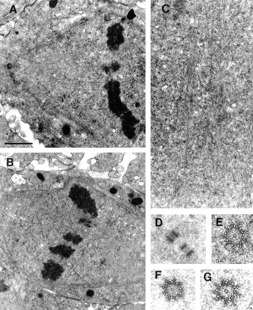
Detailed ultrastructure of MT bundles and spindle poles during mitosis. (A) Kinetochore MT bundles. (B) Interpolar MT bundles during metaphase. (C) Interpolar MT bundles during anaphase. D–G show the organization and structure of centrioles throughout mitosis. (D) Spindle poles contain two orthogonally positioned centrioles. The image shown is from the pole of a metaphase spindle. (E) During prophase, each centriole consists of nine singlet MTs around a central singlet MT. (F) by metaphase, the centrioles become much more electron dense and some of the outer MTs begin to form doublets. (G) In anaphase, doublets continue to form on the outer MTs. The standard organization of centrioles, nine outer triplets offset in a barrel shape, is never observed in Drosophila early embryos. Bar: (A) 1.3 μm; (B) 1.5 μm; (C) 1.1 μm; (D) 1.2 μm; (E–G) 0.3 μm.