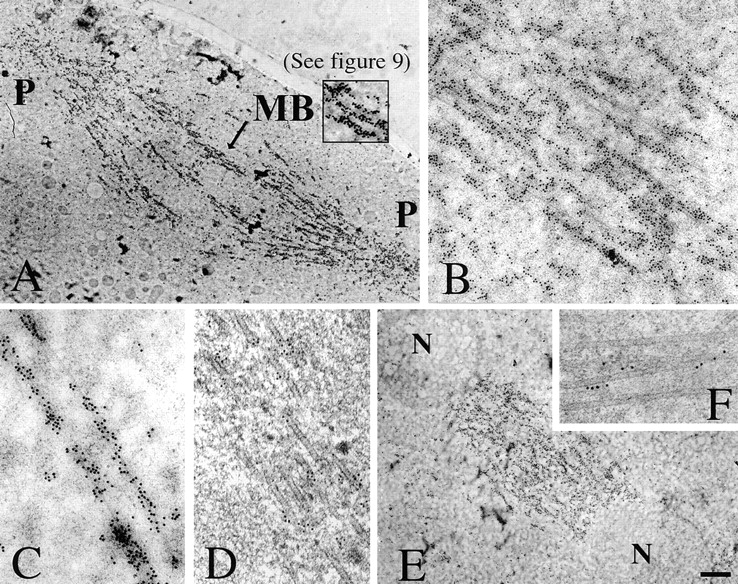
Figure 8.

Visualization of phospho-KLP61F associated with interpolar MT bundles in metaphase and anaphase spindles. EMs of thin sections of Drosophila syncytial blastoderms preserved by HPF/FS and immunostained with the anti p-BCB antibody and gold conjugated secondary antibodies. (A and E) Phospho-KLP61F staining pattern on whole metaphase and anaphase spindles, respectively, from embryos embedded in LR-White and labeled with secondary antibodies conjugated to 10 nm gold (P, spindle poles; MB, interpolar MT bundle; N, separating daughter nuclei). (A inset) Higher magnification of the interpolar bundle (MB) indicated by the arrow and shown in digitally enhanced contrast in Fig. 9. When this region is enhanced for contrast, the gold can be seen to associate with electron-dense crossbridges between microtubules (Fig. 9). (B and C) Higher magnification of the immunostaining pattern in metaphase spindle midzones. (D and F) Phospho-KLP61F immunostaining pattern on interpolar MT bundles during metaphase and anaphase B, respectively, from embryos embedded in Eponate 12/Araldite and stained with secondary antibodies conjugated to 5 nm gold to increase the precision with which the proximity of the gold to the MT can be determined. Gold particles are closely associated with the surface of MTs within these bundles. Bar: (A and E) 1.3 μm; (B) 320 nm; (C) 160 nm; (D and F) 100 nm.
