
Figure 8.
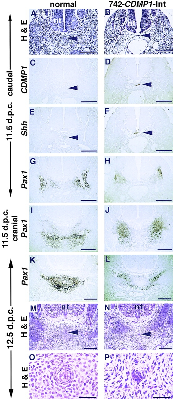
Histology and expression analysis of spine of 742- CDMP1-Int transgenic mice. (A–H) Axial semiserial sections at caudal level (in tail region) of the spine of normal (A, C, E, and G) and 742-CDMP1-Int transgenic (B, D, F, and H) embryos at 11.5 d.p.c. Staining with hematoxylin and eosin (A and B). In situ hybridization of CDMP1 (C and D), Shh (E and F), and Pax1 (G and H) antisense probes. In transgenic mice, CDMP-1 mRNA was localized in the notochord. (I and J) In situ hybridization of sections at the cranial level (thoracic region) of the spine of normal (I) and 742-CDMP1-Int transgenic (J) embryos at 11.5 d.p.c. using Pax1 antisense probe. (K–N) Axial semiserial sections of spine in the thoracic region in normal (K and M) and 742- CDMP1-Int transgenic (L and N) embryos at 12.5 d.p.c. In situ hybridization of Pax1 (K and L) and staining with hematoxylin and eosin (M and N). In normal mice, Pax1-expressing sclerotome cells migrated toward the notochord to form aggregation as the developmental stage progressed (G, I, and K). In transgenic mice, those cells remained laterally (H, J, and L). (O and P) Magnifications of the region around the notochord in M and N, respectively. In 742-CDMP1-Int transgenic mice, mesenchymal cells around the notochord were disorganized, and the sheath of the notochord was not clear. Arrowhead, notochord; nt, neural tube. Scale bars, A–N, 200 μm; O and P, 50 μm.