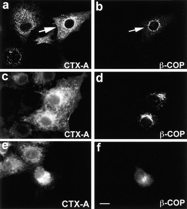
Figure 10.
Fab fragments from IgGs directed against the EAGE peptide of β-COP inhibit the translocation of CTX-A–K63 from the Golgi to the ER in Vero cells. Fab fragments were microinjected 10–15 min before starting the uptake of CTX– K63 as in Fig. 2. Incubation of the cells was continued for another 120 min. The cells were then fixed and processed for double immunofluorescence. Microinjected cells are identified by immunofluorescence of Cy2-labeled anti–β-COP Fab fragments that had been mixed with the unlabeled anti–β-COP Fab fragments at a 1:3 ratio. a, c, and e show CTX-A–K63; b, d, and f β-COP. a and b were obtained by laser scan microscopy (microinjected cell indicated by arrow); c–f were obtained by conventional immunofluorescence microscopy. Bar, 10 μm.

