Figure 5.
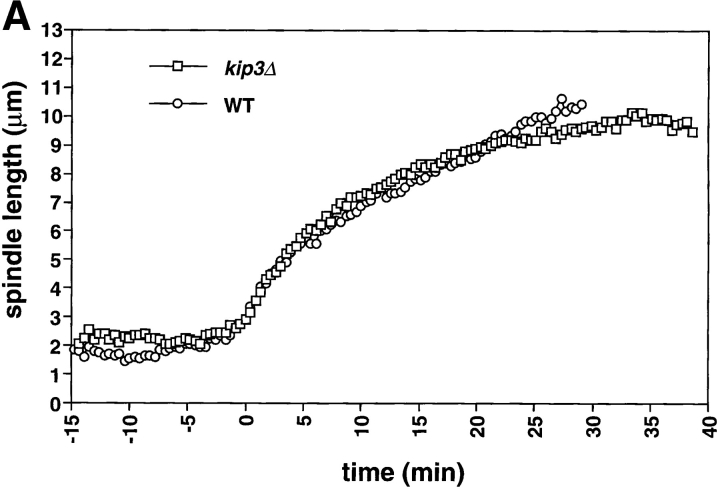
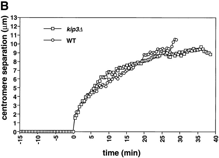
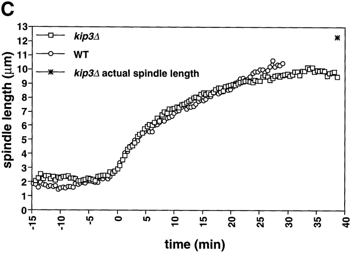
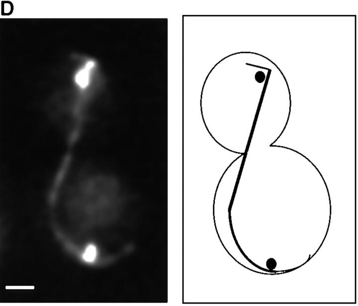
The length of the spindle and the duration of anaphase are regulated by Kip3. Cells deleted for Kip3 (AFS417) were recorded as described in Fig. 1. (A) Distance between spindle pole bodies versus time. (B) Centromere separation versus time. (C) Measurement of actual spindle length in three dimensions. Asterisk represents the total length of the spindle as compared with the distance between the spindle pole bodies. (D) Bent spindle at the end of anaphase in kip3Δ cell. Twelve 0.5-μm axial sections through a kip3Δ cell were projected onto two dimensions (left), a schematic of the cell is also shown (right). The bright dots of staining are the GFP-LacI staining of the centromeres. Bar, 2.0 μm.


